![]() Updated: 12/2/96
Updated: 12/2/96
![]() Updated: 12/2/96
Updated: 12/2/96
![]() How is SELF recognized?
How is SELF recognized?
![]() How is NONSELF (foreign) recognized?
How is NONSELF (foreign) recognized?
Clearly these are two sides of the same coin and the answer to one inevitably leads to understanding the other. Consider the DEVELOPING EMBRYO in a multicellular organism like a mammal. With immunity a multicellular organism must take into account the fact that its cellular constituents, except for identical twins, belong to a very unique gene pool of ONE. However, the fetus is not SELF, but it can not be attacked as nonself if the species is to survive. Once self recognition is achieved the multicellular organism must now differentiate between self and ALL the other NONSELF material on the planet, including its own progeny; clearly a formidable task.
As has been described previously, the problem of COMMUNICATION between biological molecules such as enzymes, their substrates, and their regulatory molecules, as well as in phage/virus binding etc. has been solved through the principle of SPECIFIC LIGAND/RECEPTOR INTERACTION. Thus the problem of differentiating between self and nonself is not one of developing a "specific recognition system", since that already exists, but how does a multicellular organism design a system for discriminating self from the millions of NONSELF substances in the environment throughout its life time? It turns out that the immune response depends upon the process of genetic recombination to solve this problem.
One final point: We CAN NOT SURVIVE without a functioning immune system. Without it, no amount of antibiotics or medical treatment can keep us alive for more than a brief time. This is painfully illustrated by the death of AIDS victims.
![]() To
learn the basic components of the immune
system
To
learn the basic components of the immune
system
![]() To
understand how the immune system works
at a fundamental level
To
understand how the immune system works
at a fundamental level
![]() To
gain an understanding of the future of immune
research and it potential impact on OUR LIVES.
To
gain an understanding of the future of immune
research and it potential impact on OUR LIVES.
![]() Women in a number of undeveloped countries put breast
milk in the infected eyes of their infants. Why might they do that?
If you were a health worker would you advise them to continue this "treatment"
or would you warn them that milk is a great medium from the growth of microbes
and advise them to stop doing this?
Women in a number of undeveloped countries put breast
milk in the infected eyes of their infants. Why might they do that?
If you were a health worker would you advise them to continue this "treatment"
or would you warn them that milk is a great medium from the growth of microbes
and advise them to stop doing this?
![]() ANTIGEN
= An antigen is anything that
ELICITS
the formation of a specific immune response. Older definitions limits the
definition of an antigen to ".....formation of an antibody.", however,
as you will learn there are two levels (duality) to the immune system.
ANTIGEN
= An antigen is anything that
ELICITS
the formation of a specific immune response. Older definitions limits the
definition of an antigen to ".....formation of an antibody.", however,
as you will learn there are two levels (duality) to the immune system.
![]() EPITOPES
= These are the PARTICULAR CHEMICAL GROUPS
on a molecule that are antigenic; that elicit a specific immune response.
EPITOPES
= These are the PARTICULAR CHEMICAL GROUPS
on a molecule that are antigenic; that elicit a specific immune response.
Click here for a view of an antibody docking with an virus epitope.
![]() ANTIBODY
= A SPECIAL GROUP OF SOLUBLE
PROTEINS that are produced in response to foreign antigens.
To view the structure of an antigen, antibody and epitope see the RasMol:Gallery
and view the section on Antibody and antigen binding. Also take a look
at the following:
ANTIBODY
= A SPECIAL GROUP OF SOLUBLE
PROTEINS that are produced in response to foreign antigens.
To view the structure of an antigen, antibody and epitope see the RasMol:Gallery
and view the section on Antibody and antigen binding. Also take a look
at the following:
Index of antibody movies. You'll have to have the right "Helper Applications" and a lot of memory to see these so take care.
This site contains the best tutorial on antibody structure I've seen. It requires the helper application Chime, Netscape 3.0 or better and a fast computer. It is fantastic, although it is advanced for Micro 101 students, if you go through this I guarantee that you'll understand what antibodies are and how they work. http://www.kumc.edu/research/medicine/biochemistry/bioc800/start.html; 11/5/96,
![]() IMMUNE
CELLS or LYPHOCYTES= These
are the VARIOUS CELLS of the specific
immunity system that respond to SPECIFIC
foreign or nonself antigens.
IMMUNE
CELLS or LYPHOCYTES= These
are the VARIOUS CELLS of the specific
immunity system that respond to SPECIFIC
foreign or nonself antigens.
Antigens are usually MACROMOLECULES
like proteins and polysaccharides; small molecules usually make POOR antigens.
Antibodies are a group of soluble, PROTEINS
that have UNIQUE BINDING
SITES on them which recognize and bind to the EPITOPES
of antigens. As previously described with enzymes, allosteric sites and
other binding site-situations, the antibody binding sites are HIGHLY
SPECIFIC. There are SEVERAL TYPES
of antibodies with a variety of different functions in the specific immune
response which will be discussed as appropriate. Figure 1 illustrates the
relationship between an antigenic molecule, its epitopes and the soluble
antibodies produced against it.
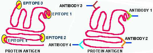
Figure 1. On the left is illustrated a folded, functional protein. It might be an enzyme, or a cell wall receptor site protein or a ribosomal protein etc. On this protein there are certain GROUPS OF ATOMS that comprise EPITOPES. These groups are defined as epitopes BECAUSE THEY ELICIT AN IMMUNE RESPONSE and for no other reason. Since each of the epitopes is a DIFFERENT and unique chemical cluster, each one of them induces a UNIQUE ANTIBODY. Each antibody will bind tightly to its particular epitope and NOT TO ANY OF THE OTHERS. Within this cartoon lies the core information one needs to understand how the immune system works. That is, if you know how one car works, you have the core information on how all cars work, only some details differ.
![]() INNATE
IMMUNITY = This can best be described as GENETIC
IMMUNITY or that immunity an organism is BORN
WITH. This type of immunity can be an immunity that applies
to the vast majority of the members of a species (SPECIES
IMMUNITY), or it can be an immunity that applies to only a certain
subgroup within a species down to a few individuals within that species.
For example, cattle suffer from the cowpox virus, but appear to have a
SPECIES IMMUNITY to the closely related smallpox viruses, whereas smallpox
is a deadly disease to humans , but cowpox is a mild localized skin infection.
Humans are susceptible to the HIV virus, but most of our related primates
are immune to HIV, but they suffer from HIV-like viruses to which we appear
to be immune. Within a species there may exist SUBGROUPS that are STATISTICALLY
immune or resistant to particular pathogens. For example, the Northern
Europeans appears to be more resistant to tuberculosis than are most Africans,
whereas Africans are naturally resistant to a variety of African diseases
that readily kill the "whites". Finally, because of the genetic variation
within every species INDIVIDUALS are resistant to some diseases, and susceptible
to other diseases. Most of you know those within your own families that
"rarely" get colds or the flu, while other family members catch one respiratory
infection after another. While there are many factors that could explain
these individual differences, one of them is that certain COMBINATIONS
OF GENES render some more resistant to the common cold viruses, whereas
others of us are very susceptible. This type of immunity has NOTHING TO
DO WITH the type of specific immunity we are discussing in this section.
INNATE
IMMUNITY = This can best be described as GENETIC
IMMUNITY or that immunity an organism is BORN
WITH. This type of immunity can be an immunity that applies
to the vast majority of the members of a species (SPECIES
IMMUNITY), or it can be an immunity that applies to only a certain
subgroup within a species down to a few individuals within that species.
For example, cattle suffer from the cowpox virus, but appear to have a
SPECIES IMMUNITY to the closely related smallpox viruses, whereas smallpox
is a deadly disease to humans , but cowpox is a mild localized skin infection.
Humans are susceptible to the HIV virus, but most of our related primates
are immune to HIV, but they suffer from HIV-like viruses to which we appear
to be immune. Within a species there may exist SUBGROUPS that are STATISTICALLY
immune or resistant to particular pathogens. For example, the Northern
Europeans appears to be more resistant to tuberculosis than are most Africans,
whereas Africans are naturally resistant to a variety of African diseases
that readily kill the "whites". Finally, because of the genetic variation
within every species INDIVIDUALS are resistant to some diseases, and susceptible
to other diseases. Most of you know those within your own families that
"rarely" get colds or the flu, while other family members catch one respiratory
infection after another. While there are many factors that could explain
these individual differences, one of them is that certain COMBINATIONS
OF GENES render some more resistant to the common cold viruses, whereas
others of us are very susceptible. This type of immunity has NOTHING TO
DO WITH the type of specific immunity we are discussing in this section.
![]() ACQUIRED
IMMUNITY = This refers to immunity that one acquires in one
of two ways, ACTIVE or PASSIVE. These are subdivided into the following
further categories:
ACQUIRED
IMMUNITY = This refers to immunity that one acquires in one
of two ways, ACTIVE or PASSIVE. These are subdivided into the following
further categories:
![]() The ACTIVE forms of immunity are generally
long lived, particularly in the case of recovery from a CLINICAL INFECTION.
Sometimes this immunity it lifelong, but in other cases it is not. Vaccinations
may induce long-lived immunity, but recent data indicate that vaccinations
may not last as long as once was hoped. For example, there is a very effective
vaccine against tetanus, but it lasts only a few years and every year hundreds
of people who have been vaccinated against this bacterium die because they
have not gotten their BOOSTER
SHOTS (vaccinations given periodically to booster the immunity
of previous vaccinations) every three to five years.
The ACTIVE forms of immunity are generally
long lived, particularly in the case of recovery from a CLINICAL INFECTION.
Sometimes this immunity it lifelong, but in other cases it is not. Vaccinations
may induce long-lived immunity, but recent data indicate that vaccinations
may not last as long as once was hoped. For example, there is a very effective
vaccine against tetanus, but it lasts only a few years and every year hundreds
of people who have been vaccinated against this bacterium die because they
have not gotten their BOOSTER
SHOTS (vaccinations given periodically to booster the immunity
of previous vaccinations) every three to five years.
Shown below are some white blood cells involved in the
immune system. Some are part of the NDS and some are components of the
specific immune system. Telling the difference between these cells is difficult,
but because their individual form (morphology) and relative numbers of
the different types are important DIAGNOSTIC
tools in disease, they are carefully studied. A variety of different
stains are used to help the medical technologist and pathologist distinguish
between the different cell types. However, many are indistinguishable morphologically
and can only be differentiated by antigenic differences.

Figure 2. This figure shows examples of two normal white blood cells (WBC). The PMN stands for polymophonuclear because they contain many nuclei (the oval dark purple structures). A PMN is a nonspecific phagocytic WBC. The cell on the right is probably a LYMPHOCYTE of some type and thus is a component of the specific immunity system.
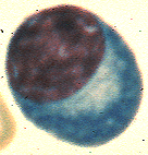
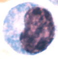

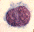
Figure 3. These four cells are various WBC. The one on the far left is a PLASMA CELL which makes antibody. Can you identify these same cells on the blood smears in lab and in the Atlas?
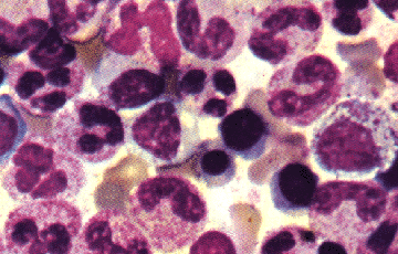
Figure 4. This is blood from a normal bone marrow. There are a variety of cells present in various stages of development or maturity, making it very difficult to accurately distinguish the types.
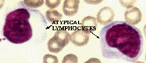
Figure 5. This is blood from a patient with infectious mono or the "kissing disease", which is common among college students for some unknown reason. There is a theory circulating that people actually enjoy the process of catching this disease. The atypical appearance of the lymphocytes is diagnostic of the disease.
The steps in the immune system development are:
![]() Stem cells, which are the PARENT CELLS
of all immune cells, enter the liver of the fetus and develop to a point
there.
Stem cells, which are the PARENT CELLS
of all immune cells, enter the liver of the fetus and develop to a point
there.
![]() From the liver some stem cells move into the bone marrow (the center of
the bones) where they differentiate into B CELLS
and NATURAL KILLER CELLS.
From the liver some stem cells move into the bone marrow (the center of
the bones) where they differentiate into B CELLS
and NATURAL KILLER CELLS.
![]() Other stem cells move from the liver into the thymus gland located in the
middle of your chest.
Other stem cells move from the liver into the thymus gland located in the
middle of your chest.
![]() The thymic stem cells differentiate in a variety of T
cells.
The thymic stem cells differentiate in a variety of T
cells.
![]() Other stem cells go on to differentiate into other blood cell lines such
as macrophages.
Other stem cells go on to differentiate into other blood cell lines such
as macrophages.
Immunologists are making headway in unraveling the complexities of these various differentiation's, but the differentiation process is extremely complex and subtle. From my perspective of >40 years in microbiology I have observed tremendous progress in the area of immunology. However, my guess would be that we are not even half way to a full understanding of the entire system. I am optimistic that the immune system will be completely understood in your lifetimes.
The immune system is spread throughout the entire body and includes the following (a partial listing):
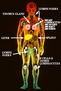
Figure 6. This figure shows the location in the body of various components of the nonspecific and specific immune systems. The B cells and a variety of other lymphatic cells are made in the bone marrow. The lymph nodes contain the macrophages, B cells and T cells, which is why your lymph glands swell up and become tender to the touch when you have an infection. The thymus gland is the gland where the differentiation of the T cells occurs. Other macrophages, monocytes and phagocytes reside in the liver, spleen and lungs. Special immune cells have been found in the brain, in the skin and in the cells lining the intestine. Breast milk contains a variety of the mother's white blood cells that kill microbes in the infant's gut and stimulate the development of the infant's immune system as well as antibodies and 10 other microbial inhibitors (Sci. Am. Dec. 1995)
![]() Instead of thinking
that the immune system had to be INSTRUCTED AHEAD
OF TIME as to which antibodies would be required throughout
a life time, clearly an impossible task, N.K. JERNE suggested that the
immune system was SELECTIVE rather
than instructive. That the immune system RANDOMLY
made billions of different SPECIFIC-EPITOPE-BINDING
ANTIBODIES and then let the antigens that accidentally stumbled
into the host chose or select which antibodies would be produced in quantities
large enough to be protective. In a sense this is just another twist on
the "survival of the fittest" process in EVOLUTION.
Burnet in Australia and Talmage in CO then hypothesized that antibodies
SIT
ON THE SURFACE of lymphocytes
and that each lymphocyte manufactures only a SINGLE
ANTIBODY (which recognizes and binds to only a SINGLE
EPITOPE). This theory has been shown to be essentially correct
by a number of brilliant experimentalists.
Instead of thinking
that the immune system had to be INSTRUCTED AHEAD
OF TIME as to which antibodies would be required throughout
a life time, clearly an impossible task, N.K. JERNE suggested that the
immune system was SELECTIVE rather
than instructive. That the immune system RANDOMLY
made billions of different SPECIFIC-EPITOPE-BINDING
ANTIBODIES and then let the antigens that accidentally stumbled
into the host chose or select which antibodies would be produced in quantities
large enough to be protective. In a sense this is just another twist on
the "survival of the fittest" process in EVOLUTION.
Burnet in Australia and Talmage in CO then hypothesized that antibodies
SIT
ON THE SURFACE of lymphocytes
and that each lymphocyte manufactures only a SINGLE
ANTIBODY (which recognizes and binds to only a SINGLE
EPITOPE). This theory has been shown to be essentially correct
by a number of brilliant experimentalists.
![]() 2.
The B cells that produce self antibodies are DESTROYED,
leaving only lines or CLONES of B cells
that produce random antibodies to foreign epitopes.
2.
The B cells that produce self antibodies are DESTROYED,
leaving only lines or CLONES of B cells
that produce random antibodies to foreign epitopes.
![]() 3.When
a particular foreign epitope (say antigen #2,025)
appears in the host's body it is PROCESSED
by lymphocytic cells of the NDS This sets off a series of events that eventually
acts on a small population of randomly-produced B cells that happen to
have on their surface, antibody (#2,025)
which binds to ANTIGEN #2,025.
3.When
a particular foreign epitope (say antigen #2,025)
appears in the host's body it is PROCESSED
by lymphocytic cells of the NDS This sets off a series of events that eventually
acts on a small population of randomly-produced B cells that happen to
have on their surface, antibody (#2,025)
which binds to ANTIGEN #2,025.
![]() 4.
These events trigger a RAPID PROLIFERATION
of that PARTICULAR B cell population
(#2,025), producing a large number
of clones. These B cell-clones differentiate into PLASMA
CELLS (Fig. 3) which are ANTIBODY-PRODUCING-FACTORIES
that spew out prodigious quantities of the ONE #2,025
ANTIBODY
that will bind to the specific antigen (epitope) that stimulated
it.
4.
These events trigger a RAPID PROLIFERATION
of that PARTICULAR B cell population
(#2,025), producing a large number
of clones. These B cell-clones differentiate into PLASMA
CELLS (Fig. 3) which are ANTIBODY-PRODUCING-FACTORIES
that spew out prodigious quantities of the ONE #2,025
ANTIBODY
that will bind to the specific antigen (epitope) that stimulated
it.
![]() 5.
The specific antibody floods through the host and wherever it binds to
its epitope it MARKS IT FOR ATTACK
and destruction by the appropriate cells and associated components of the
immune system.
5.
The specific antibody floods through the host and wherever it binds to
its epitope it MARKS IT FOR ATTACK
and destruction by the appropriate cells and associated components of the
immune system.
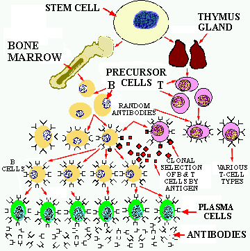
Figure 7. The process of B & T cell differentiation and CLONAL SELECTION. The parental STEM cells migrate to the bone marrow and to the thymus gland where they differentiate into B and T cells which make random epitope binding proteins. When a foreign epitope binds to the appropriate site on the B & T cells, they replicate into clones that, in the case of the B cells differentiate into PLASMA cells that produce prodigious quantities of specific antibodies. The T cell clones further differentiate into several different T cell types with specific functions.
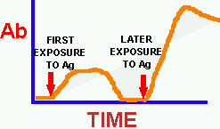
Figure 8. The response to an antigen (Ag) in terms of the production of a specific antibody over time. Initially the levels of each unique antibody are extremely low, however as soon as the stimulation events occur (Fig. 7) and the plasma cell clone begins producing antibodies the TITER (concentration or amount) of the unique antibody begins to rise. It takes about 2 weeks for the Ab level to peak. Once the foreign antigen is removed, antibody production slowly returns to a low level, however MEMORY PLASMA CELLS remain in the system. When the original antigen again appears in the host these memory cells respond rapidly and produce even higher levels of antibodies. This "REMEMBERING RESPONSE" is why we remain immune to many diseases for a long time. The secondary exposure to the antigen may be natural or it may be artificial in the case of BOOSTER vaccinations. As parents we are responsible for seeing to it that our children are initially vaccinated and that their booster shots are given at the appropriate ages.
There are five different types of antibodies, however
in this course we will discuss only the most common one, IgG,
in detail. However, note that the other 4 types physically resemble the
basic structure of IgG. IgG does most of the humoral immune work. The figure
below shows the physical structure of the IgG molecule.
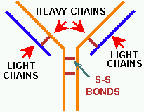
Figure 9. The IgG molecule. IgG is composed of two protein subunits, a LIGHT and a HEAVY CHAIN named appropriately according to their relative sizes. The various chains are bonded together to form the IgG molecule with disulfide bonds (S-S bonds). Molecular antibody model; note the two arms & the heavy 'n light chains.
The Y-shaped structure is real as electron microscopic pictures show. However, even before they viewed IgG in an EM immunologist had discerned its basic shape. They knew that each antibody had to have two equivalent binding sites for its specific epitope. It turns out that those two binding sites are located at the end of the short arms of the Y (Fig. 10).
The IgG molecule is further divided into CONSTANT
and VARIABLE REGIONS OR DOMAINS. The
constant regions have mostly the SAME amino acid sequence in all IgG molecules
(we won't discuss the differences here), whereas the amino acid sequences
in the variable regions are DIFFERENCE
for each unique antibody produced by a clone of plasma cells. The amino
acid sequence in the variable domains are such that they tightly bind to
particular epitopes. Thus they show the same LOCK-KEY
relationship as do enzymes/substrates and enzymes/allosteric molecules
and viruses/target
cell receptors.
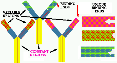
Figure 10. Three unique antibody IgG molecules. The base of the "Y" and part of each arms are called the CONSTANT REGIONS because their amino acid sequence tends to be very similar in all IgG molecules. The variable regions are at the end of the arms and their amino acid sequence is very different for each IgG molecule. These variable regions fold so as to bind to specific epitopes or antigens; the unique binding sites are shown in their respective three variable regions on the right.
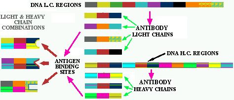
Figure 11. Random Ab formation. Each of the colored squares in the light chain (L.C.) and heavy chain (H.C.) regions represent a GENE FRAGMENT. If three of these fragments are required to make one gene for the VARIABLE REGION a large number of combinations are possible, some of which are shown below each cluster of fragments. Then on the far left are several examples of combinations between the variable light and heavy chain genes that form the variable arms of IgG. As you can see from the limited numbers of color bars used in the illustration many different combinations could be formed.
Click here to see a series of views of antibody molecules.
Click
here to see the 5 types of antibody molecules; note the similarities
and differences between them.
NEUTRALIZATION = When the antigen is a soluble toxin, the addition of an Ab against it will usually render the toxin INEFFECTIVE, that is it NEUTRALIZES it. Such neutralized toxins are called TOXOIDS and can be used as vaccines. For example, if you were suspected of suffering from either tetanus or botulism poisoning the treatment would involve giving you a shot of the appropriate antitoxin, which is a common name for the Ab against a toxin. The antitoxin circulates through your body and binds and neutralizes any toxin it contacts.
PRECIPITATION = Under the proper conditions a soluble antigen can be precipitated in the presence of its Ab because of the of antigen-antibody net-work that forms gets large enough to form masses that SETTLE OUT (precipitate) on their own.
AGGLUTINATION
= When the antigen is a large PARTICLE,
like a whole bacterium or a RBC, the addition of its Ab will form an Ab-antigen
net work that causes the particles to CLUMP IN LARGE MASSES like milk coagulating
when it spoils. This agglutination is easy to see and is useful for diagnostic
purposes. For example, if you want to see if a person is making Ab against
a particular bacterium, mix the person's serum with the suspected bacterium;
if the bacteria clump into large globs it means that Ab are present. Both
precipitation and agglutination are illustrated below.
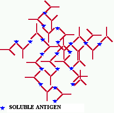
Figure 12. Antibody/antigen complex forming a larger complex. These nets can grow so large that they become insoluble and visible to the eye. The network forms because of the dual-binding characteristic of the antibody which allows it to attach to two different antigen molecules at the same time.
 We
formally used the agglutination test to determine the blood type of students
in Micro 101, but because of the danger of AIDS it is no longer considered
safe to do this test under lab conditions.
Does this raise a concern in your mind about having sex with someone whose HIV status you don't know if it is considered unsafe to test blood types in a controlled laboratory setting?
Click here to see an illustration of the interaction between a T-Helper cell and an antigen-presenting cell.

Figure 13. Activation of & killing by Tc killer cells of cells displaying a unique surface antigen. Note the virus particles in the cell on the right and the presence of unique viral proteins on its surface to which the Tc cells bind.
Other T cell types exist and probably more types will be found. The above is an incomplete and simplified explanation of what is currently known about the immune system. Some of it will undoubtedly be modified as new facts come to light and we will surely find that it is even more complex and subtle than previously imagined. It's like human relationships which usually start out simple, but the become more complex as time goes on.
Since you've been paying attention, you should suspect by now that autoimmune disease is complex. In many autoimmune illnesses, genetic factors are present. For example, identical twins have a high chance of suffering from the same AD. The causes of AD are virtually unknown. A significant amount of data indicates that infections can trigger them, or they can be provoked simply by an injury or stress. There are some hopeful signs of treatment for some of these AD, but much more needs to be learned about them.
![]() An initial exposure of the immune system to an ALLERGEN.
At this time there are NO SYMPTOMS as the immune system must synthesize
the IgE.
An initial exposure of the immune system to an ALLERGEN.
At this time there are NO SYMPTOMS as the immune system must synthesize
the IgE.
![]() On subsequent exposures to the allergen it binds to IgE molecules that
are located on the surface of MAST CELLS.
On subsequent exposures to the allergen it binds to IgE molecules that
are located on the surface of MAST CELLS.
![]() This induces a CASCADE of events that
cause the mast cells to release chemicals present in granules in the mast
cells.
This induces a CASCADE of events that
cause the mast cells to release chemicals present in granules in the mast
cells.
![]() These chemicals include histamines, leukotrienes and prostaglandins, which
in turn INDUCE THE VARIOUS SYMPTOMS typical of an allergic response.
These chemicals include histamines, leukotrienes and prostaglandins, which
in turn INDUCE THE VARIOUS SYMPTOMS typical of an allergic response.
This entire process can takes JUST
SECONDS, thus explaining the suddenness with which allergic
and hyptersensitive reactions can occur. Allergies include a WIDE VARIETY
of diseases. For example chronic allergic rhinitis (runny nose, stuffed
up sinuses) is commonly caused by the feces of the COMMON
HOUSE LOUSE (no! not your mate or the cat) which lives in our
homes in rugs and on furniture. Seasonal allergies are often caused by
pollens or mold spores in the air. Asthma effects approximately 5 to 10%
of children, but another 5 to 10% acquire asthma in adulthood and others
become afflicted in their 80s. There are numerous forms of asthma, only
some of which may involve IgE-mediated activity.
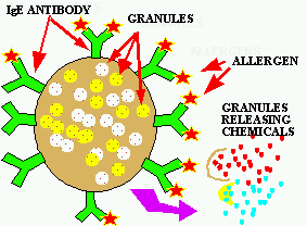
Figure 14. Allergic response. A MAST cell coated with specific IgE antibody to an allergen reacts with the allergen, triggering the rapid release of the "chemical containing granules" within the MAST cell. These granules burst and release these potent chemicals which bring on the allergy attack.
![]() After
the end of the second world war some American service men in Japan suffered
from painful and itchy blisters developing on their elbows and in a ring
around their buns. Was this a subtle form of chemical warfare developed
by the Japanese as retaliation for losing? No, it turned out that a common
Japanese wood used to make toilet seats and bar tables contains a chemical
that was very similar to poison ivy allergen and the Americans were reacting
to it because of their exposure to poison ivy in the US.
After
the end of the second world war some American service men in Japan suffered
from painful and itchy blisters developing on their elbows and in a ring
around their buns. Was this a subtle form of chemical warfare developed
by the Japanese as retaliation for losing? No, it turned out that a common
Japanese wood used to make toilet seats and bar tables contains a chemical
that was very similar to poison ivy allergen and the Americans were reacting
to it because of their exposure to poison ivy in the US.
There are many myths and much misunderstanding about allergies. Much money is spent on testing for allergies and for allergy treatments, but RIGOROUS PROOF is often lacking both for allergies or for the efficacy of the, usually expensive, treatments. The role of industrial pollutants in producing allergies is not clear, but much data suggests a relationship between air quality, asthma and other respiratory difficulties. Much work remains to be done before this relationship is resolved. There are treatments for eliminating the sensitivity to specific allergens, but they generally required a lifetime commitment to the treatment. Before embarking on a long series of expensive, sometimes painful injections to treat your allergies, it is a good idea to get a second opinion and to explore alternative treatments. For example, one of the most common causes of household allergies is the feces of a louse that lives in all our homes. By vacuuming more frequently using special bags that TRAP the LOUSE FECES you may decrease the frequency and severity of your allergy attacks. Air purification and filtration systems may offer a viable option to shots. Also wearing protective particle masks when engaging in activities that are likely to expose you to allergens (e.g. cleaning the house), and changing clothes/bathing immediately after, may prevent allergy attacks.
![]() What do you think? What percentage of YOUR INCOME are you prepared to spend
on research like this (as taxes)? Do you think it would be better to let
people carrying these "bad" genes die before having children and passing
them on for future generations to care for? What role does society have
in treating people with "bad" genes? Who decides what a "bad gene" is?
What do you think? What percentage of YOUR INCOME are you prepared to spend
on research like this (as taxes)? Do you think it would be better to let
people carrying these "bad" genes die before having children and passing
them on for future generations to care for? What role does society have
in treating people with "bad" genes? Who decides what a "bad gene" is?
Collection of antibody images.
Another course on the immune system. Very good pictures and complete explanations; might even be better than my descriptions (gasp!!!).
SCIENCE HALL, ROOM 440CA
PHONE: 509-335-5108
FAX: 509-335-1907
E-mail address:
hurlbert@wsu.edu hurlbert@pullman.com
OFFICE HOURS: M,W 1:30 to 3:30 PM.